
Wavefront là gì ?công nghệ mặt sóng tối tân nhất của Israen trong kiểm tra mắt
Công nghệ mặt sóng được phát triển cho LASIK tùy chỉnh có thể sớm được các bác sĩ nhãn khoa sử dụng thường xuyên để chẩn đoán tốt hơn các vấn đề về thị lực…


Công nghệ mặt sóng được phát triển cho LASIK tùy chỉnh có thể sớm được các bác sĩ nhãn khoa sử dụng thường xuyên để chẩn đoán tốt hơn các vấn đề về thị lực trong các kỳ thi mắt, có thể khiến biểu đồ mắt quen thuộc trở nên lỗi thời.
Hầu hết mọi người đã khám mắt bằng một thiết bị gọi là phoropter , trong đó có nhiều thấu kính có độ phóng đại khác nhau. Bác sĩ nhãn khoa hoặc bác sĩ đo thị lực sẽ thay đổi thấu kính trước mắt bạn, hỏi xem thấu kính nào tạo ra hình ảnh tốt nhất.
Với cách tiếp cận thông thường này, thông tin bạn cung cấp cho bác sĩ nhãn khoa rất chủ quan, dựa nhiều hơn vào những gì bạn nghĩ mình nhìn thấy thay vì những gì bạn thực sự nhìn thấy. Nhưng phép đo mặt sóng là khách quan, vì lỗi thị lực có thể được xác định tự động theo cáh sóng ánh sáng truyền qua mắt .
Một ngày nào đó, những phép đo mặt sóng chi tiết này có thể thay thế đơn thuốc kính mắt hoặc kính p tròng thông thường , vốn chỉ mô tả các vấn đề về thị lực theo độ cận thị, viễn thị và loạn thị của mắt .
Cũng giống như LASIK tùy chỉnh (hay "hướng dẫn theo mặt sóng") có khả năng mang lại tầm nhìn sắc nét hơn so với LASIK thông thường, kính và kính áp tròng được sản xuất bằng công nghệ tiên tiến này cũng có thể mang lại tầm nhìn rõ nét hơn so với các loại kính thông thường.
Để biết thêm về sự khác biệt giữa đơn thuốc kính mắt thông thường và đơn thuốc kính mắt hướng sóng, hãy đọc: Tròng kính mắt có độ nét cao mang lại tầm nhìn sắc nét hơn như thế nào
Khám mắt mặt sóng là gì?
Vậy thì "mặt trận sóng" thực chất có nghĩa là gì?
Hãy tưởng tượng ánh sáng di chuyển như một chùm tia sáng. Nếu bạn vẽ các đường vuông góc với các đầu của một chùm tia sáng, bạn sẽ có được cái gọi là bản đồ mặt sóng. Trong một mắt có thị lực hoàn hảo, mặt sóng hoàn toàn phẳng, nhưng mặt sóng của một mắt không hoàn hảo thì không đều.
Các loại biến dạng mà mặt trận sóng này thu được khi truyền qua mắt cung cấp thông tin có giá trị về các lỗi thị lực và cách khắc phục chúng.
Công nghệ mặt sóng (quang sai) là gì?
Aberrometry đo cách mặt sóng ánh sáng đi qua giác mạc và thủy tinh thể , là các thành phần khúc xạ (tập trung ánh sáng) của mắt. Sự biến dạng xảy ra khi ánh sáng đi qua mắt được gọi là quang sai, biểu thị các lỗi thị lực cụ thể.Công nghệ mặt sóng, hay còn gọi là quang sai, chẩn đoán cả lỗi thị lực bậc thấp và bậc cao biểu thị bằng cách mắt khúc xạ hoặc hội tụ ánh sáng.Quang sai bậc cao là lỗi thị lực phức tạp hơn, trong khi quang sai bậc thấp là lỗi thị lực phổ biến hơn như cận thị , viễn thị và loạn thị.
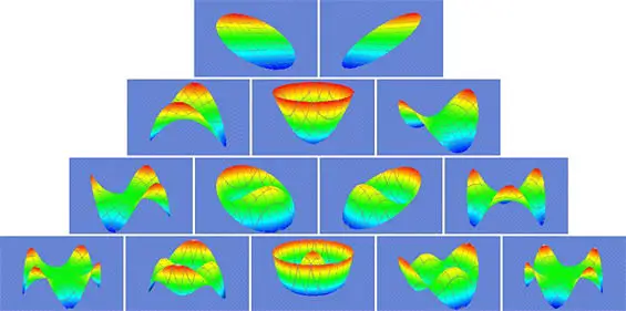
hình dạng quang sai của mắt
Mặt trận sóng và quang sai bậc cao
Trước đây, với các phương pháp kiểm tra mắt thông thường, chỉ có thể chẩn đoán và điều trị các lỗi thị lực bậc thấp. Các quang sai bậc cao như coma, trifoil và cầu sai phần lớn bị các chuyên gia chăm sóc mắt bỏ qua vì tác động của chúng đối với thị lực được cho là không đáng kể vào thời điểm đó và vì không có phương tiện khả thi nào để xác định hoặc sửa chúng một cách chính xác.
Hiện nay, khi quang sai bậc cao có thể được xác định chính xác bằng công nghệ mặt sóng và được hiệu chỉnh bằng các loại kính mới, kính áp tròng, kính nội nhãn và phẫu thuật khúc xạ, chúng đã trở thành những yếu tố quan trọng hơn trong các kỳ thi mắt.
Các quang sai bậc cao bắt đầu nhận được nhiều sự chú ý hơn vì chúng được xác định là nguồn gây ra các tác dụng phụ về thị giác sau phẫu thuật LASIK và các loại phẫu thuật khúc xạ khác — gây ra hiện tượng quầng sáng, hình ảnh ma và các triệu chứng thị giác khác.
Các tia laser dẫn hướng mặt sóng mới được sử dụng trong phẫu thuật điều chỉnh thị lực có thể làm giảm một số quang sai bậc cao, từ đó cải thiện hiệu suất thị giác, đặc biệt là đối với các hoạt động thiếu sáng như lái xe vào ban đêm.
Phép đo mặt sóng được thực hiện như thế nào?
Chẩn đoán mặt sóng của mắt được thực hiện bằng máy đo quang sai .Khám mắt theo phương pháp truyền thống sử dụng máy đo công nghệ với thấu kính thử nghiệm có thể mất nhiều thời gian và phụ thuộc vào phản hồi chủ quan của bệnh nhân. Kiểm tra mắt mặt sóng tự động và khách quan đo các lỗi thị lực chỉ trong vài giây và chi tiết hơn nhiều.
Với hầu hết các máy đo quang sai, bạn đặt cằm lên một giá đỡ cằm. Tiếp theo, bạn được yêu cầu nhìn vào thiết bị và tập trung mắt vào một điểm sáng. Sau vài giây im lặng (không cần phản hồi), phép đo mặt sóng hoàn tất và bản đồ mặt sóng của mắt bạn sẽ được in ra cho chuyên gia chăm sóc mắt của bạn.
Nhìn chung, máy đo quang sai sử dụng quy trình ba bước:
1.Vì mặt trận sóng đi qua lỗ mở ở phía trước mắt (đồng tử), nên đường kính đồng tử của bạn được đo. Phép đo này được sử dụng để suy ra hình dạng mặt trận sóng tham chiếu đại diện cho một con mắt hoàn hảo về mặt lý thuyết có kích thước đồng tử giống hệt như mắt của bạn.
2.Một chùm ánh sáng được chiếu vào mắt bạn và được phản xạ trở lại bởi võng mạc. Mặt trận sóng của ánh sáng phản xạ này được máy đo quang sai bắt được. Vì không có mắt nào là hoàn hảo về mặt quang học, tất cả các mặt trận sóng như vậy sẽ chứa ít nhất một số biến dạng.
3.Bản đồ quang sai của mắt được tạo ra bằng cách so sánh hình dạng của mặt trận sóng thu được với mặt trận sóng tham chiếu được lập trình sẵn, đo tất cả các điểm khác biệt giữa hai mặt trận. Sau đó, bản đồ mặt trận sóng của mắt bạn, đôi khi được gọi là "dấu vân tay quang học" vì không có hai mắt nào tạo ra cùng một bản đồ chính xác, được tạo ra.
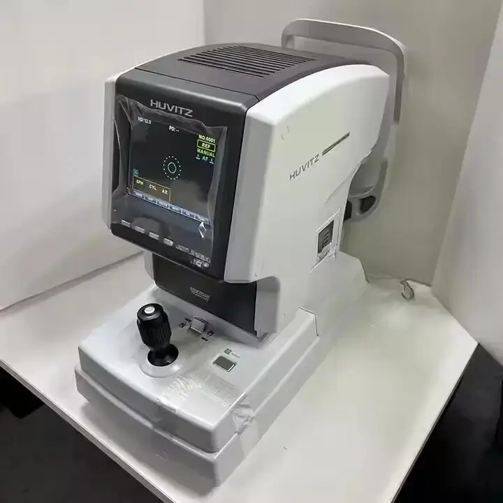
Khám mắt theo phương pháp truyền thống sử dụng máy đo với thấu kính thử nghiệm có thể mất nhiều thời gian và phụ thuộc vào phản hồi chủ quan của bệnh nhân
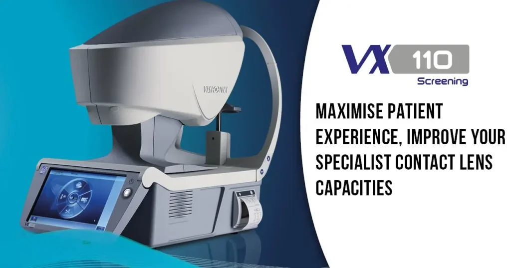
Kiểm tra mắt mặt sóng tự động và khách quan đo các lỗi thị lực chỉ trong vài giây và chi tiết hơn nhiều
Bản đồ mặt sóng của mắt bạn có ý nghĩa gì?
Khi bạn bắt đầu diễn giải kết quả bản đồ mặt sóng của mắt, hãy nhớ rằng hình dạng tham chiếu được sử dụng để so sánh là hình phẳng hoặc hai chiều.


Mặc dù không có mắt nào là hoàn hảo, bản đồ mặt sóng của mắt hoàn hảo về mặt lý thuyết là một mặt phẳng. Lưu ý cách hình dạng ba chiều này biểu diễn lỗi thị lực thay đổi thành gần như mặt phẳng sau khi mắt được điều chỉnh bằng LASIK hướng dẫn mặt sóng.
Mặt phẳng tròn này về mặt lý thuyết tượng trưng cho một mắt hoàn hảo ( emmetropic ) khớp chính xác với đường kính đồng tử mắt của bạn.
Bản đồ mặt sóng ba chiều thực tế của mắt bạn được tạo ra thông qua việc so sánh với bản đồ mặt sóng phẳng hoàn hảo về mặt lý thuyết này. Sự biến dạng ba chiều xảy ra trong bản đồ mặt sóng thực tế dựa trên cách các tia sáng riêng lẻ bị thay đổi khi chúng đi qua mắt.
Các khiếm khuyết ở giác mạc và thủy tinh thể có thể làm thay đổi đường đi của tia sáng bằng cách khiến chúng tăng tốc, chậm lại hoặc bẻ cong không đúng cách (khúc xạ).
Các vấn đề về mắt khác cũng có thể gây ra quang sai bậc cao, bao gồm khô mắt làm thay đổi màng nước mắt của mắt, cũng giúp khúc xạ các tia sáng. Ngay cả đục thủy tinh thể làm mờ thấu kính tự nhiên của mắt hoặc sẹo trên bề mặt mắt do phẫu thuật mắt, bệnh tật hoặc chấn thương cũng có thể gây ra quang sai bậc cao và các vấn đề về hội tụ.
Dựa trên nhiều kiểu mẫu khác nhau có thể xuất hiện trên mặt trận sóng, hơn 60 hình dạng quang sai khác nhau đã được xác định và phân loại là lỗi thị giác.
Đo mặt sóng
Các phép đo mặt sóng xác định lỗi thị lực có thể được tính theo nhiều cách khác nhau. Hiện tại, phương pháp được lựa chọn là một biểu thức toán học gọi là đa thức Zernike.
Được nhà vật lý người Hà Lan Fritz Zernike định nghĩa vào năm 1934, đa thức này rất phù hợp với nhiệm vụ này vì nó được thiết kế để đáp ứng các yêu cầu của một đường tròn.
Mỗi đa thức Zernike mô tả quang sai tồn tại tại một điểm cụ thể trên mặt sóng ánh sáng, sau khi nó đi qua mắt. Tổng của các đa thức Zernike bằng mô tả đầy đủ về tất cả các quang sai hoặc tổng lỗi khúc xạ trong một mắt nhất định.
Phát hiện này được đưa ra dưới dạng đơn thuốc Zernike và bản đồ địa hình, bản vẽ chi tiết về hình dạng của mặt sóng quang sai. Cùng nhau, chúng tạo nên bản đồ quang sai hoặc "dấu vân tay quang học" độc đáo xác định chi tiết các lỗi thị lực của mắt.
Bản đồ mặt sóng của bạn được sử dụng như thế nào
Bản đồ mặt sóng là mô tả đầy đủ, chính xác về mọi quang sai ảnh hưởng đến mắt bạn. Bác sĩ nhãn khoa sử dụng bản đồ mặt sóng của bạn như một bản thiết kế để thiết kế riêng phương pháp điều chỉnh thị lực cho bạn, cho dù là phẫu thuật khúc xạ như LASIK, thấu kính nội nhãn, kính áp tròng hay kính đeo mắt.
Hãy nhớ rằng bạn không bao giờ có thể đạt được mặt sóng hoàn hảo, vì không có mắt nào là hoàn hảo. Ngay cả khi bạn được chẩn đoán mắc một số quang sai bậc cao nhất định, điều này không nhất thiết là nguyên nhân gây lo ngại, trừ khi mức độ quang sai gây ra các vấn đề đáng kể về thị lực như khó nhìn vào ban đêm.

Hiệu suất thị giác của hệ thống quang học (mắt) của bệnh nhân dựa trên các chỉ số đo quang sai, chia thành các thành phần giác mạc và bên trong.
Tài liệu tham khảo
-
Maeda, N., 2009. “Ứng dụng lâm sàng của phép đo quang sai mặt sóng - một bản tóm tắt.” Nhãn khoa lâm sàng và thực nghiệm, 37(1): tr.118-129.
-
Salés, C. và E. Manche (2015). “So sánh quang sai mắt được đo bằng máy đo quang sai Hartmann-Shack dựa trên Fourier và máy đo quang sai Tscherning dựa trên Zernike trước và sau khi phẫu thuật cắt bỏ giác mạc bằng laser tại chỗ.” J Cataract Refract Surg 41(1): 1820-1825.
-
Ohlendorf, A., et al., 2020. “Nâng cao quy trình làm việc kỹ thuật số để đo lỗi khúc xạ.” Tạp chí Y học lâm sàng, 9(1): trang 2205.
-
Jinabhai, A. (2019). “Các biện pháp điều chỉnh kiểm soát quang sai tùy chỉnh cho bệnh nhân bị giác mạc hình nón sử dụng kính áp tròng.” Clin Exp Optom 103(1): 31-43.
-
Kumar, P., et al. (2019). “Hiệu suất thị giác và chất lượng quang học có khác nhau giữa các phương thức điều chỉnh kính áp tròng khác nhau ở bệnh keratoconus không?” Contact Lens and Anterior Eye, S1367-0484(20):30052-30057.
-
Hoy, L., 2019. “Giải mã quá trình định hình lại giác mạc bằng Ortho-K.” Bài giảng cho CCLSA Victoria.
Để lại bình luận
Lưu ý: Bình luận của bạn sẽ được kiểm duyệt trước khi hiển thị. Vui lòng giữ nội dung lịch sự và phù hợp.